Acute Management of Ankle Injuries: What You Should Do in the First 72 Hours

Ankle injuries are one of the most common musculoskeletal injuries, affecting everyone from elite athletes to weekend walkers. Whether you've rolled your ankle playing sport, stepping off a curb, or hiking on uneven ground, how you manage the injury in the first few days can have a significant impact on your recovery.

The good news? Most ankle injuries recover well with the right treatment and rehabilitation.

What Happens When You Roll Your Ankle?

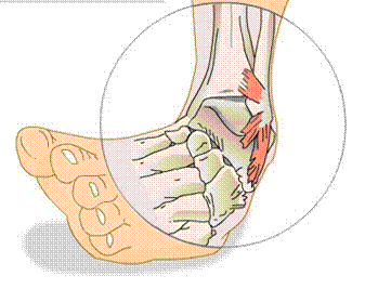
The majority of ankle injuries involve a lateral ankle sprain, where the foot rolls inward (inversion), overstretching or tearing the ligaments on the outside of the ankle.

Common symptoms include:

  • Pain around the outside of the ankle

  • Swelling

  • Bruising

  • Difficulty walking or bearing weight

  • A feeling of instability or "giving way"

The severity can range from a mild ligament stretch (Grade I) to a complete ligament tear (Grade III).

What Should You Do Immediately After the Injury?

Current best practice recommends following the PEACE & LOVE principles:

PEACE (First 1–3 Days)

P – Protect
Reduce activities that increase pain during the first 1–3 days. You don't necessarily need complete rest, but avoid movements that significantly aggravate the injury.

E – Elevate
When possible, elevate your ankle above heart level to help reduce swelling.

A – Avoid Anti-inflammatory Medication
While anti-inflammatory medications may reduce pain, inflammation is an important part of tissue healing. They should not be used routinely in the early stages unless recommended by your healthcare provider.

C – Compress
Use a compression bandage or ankle sleeve to help manage swelling and provide support.

E – Educate
Avoid relying solely on passive treatments. Active rehabilitation is the key to achieving the best long-term outcomes.

LOVE (After the First Few Days)

As pain begins to settle, focus on gradually returning to movement.

L – Load
Begin gentle weight-bearing and movement as tolerated. Pain can be used as a guide—some discomfort is acceptable, but pain should not progressively worsen.

O – Optimism
Recovery is influenced by both physical and psychological factors. Staying positive and understanding the healing process can improve outcomes.

V – Vascularisation
Introduce low-impact cardiovascular exercise, such as cycling or swimming, as symptoms allow. This promotes circulation and supports healing.

E – Exercise
Progressive rehabilitation restores:

  • Strength

  • Balance

  • Mobility

  • Coordination

  • Confidence

These are essential for preventing recurrent ankle sprains.

Should You Ice Your Ankle?

Ice can help reduce pain in the short term and may make the ankle feel more comfortable. However, research suggests that icing does not speed up healing and should be used primarily for pain relief rather than as a treatment to accelerate recovery.

If using ice:

  • Apply for 10–15 minutes at a time.

  • Wrap the ice pack in a towel.

  • Allow the skin to return to normal temperature before reapplying.

When Should You See a Physiotherapist?

Not every ankle sprain requires an X-ray, but every significant ankle injury benefits from an accurate assessment.

You should seek assessment if:

  • You are unable to walk four steps immediately after the injury or the following day.

  • Swelling or bruising is severe.

  • Pain persists beyond a few days.

  • Your ankle feels unstable.

  • You continue to sprain the same ankle repeatedly.

  • You're aiming to return to sport or an active lifestyle safely.

A physiotherapist can determine the severity of the injury, identify whether other structures (such as tendons or the syndesmosis) may be involved, and develop a rehabilitation program tailored to your goals.

Why Rehabilitation Matters

Many people stop treatment once the pain settles. Unfortunately, pain often improves before the ligaments, muscles, and balance systems have fully recovered.

Without proper rehabilitation, individuals are more likely to experience:

  • Recurrent ankle sprains

  • Chronic ankle instability

  • Persistent pain

  • Reduced sporting performance

  • Long-term ankle problems

Research shows that structured exercise therapy significantly reduces the risk of future ankle sprains.

The Bottom Line

Most ankle sprains recover well, but the first few days are important. Protect the ankle, manage swelling, gradually return to movement, and begin a structured rehabilitation program as early as appropriate.

If you're unsure about the severity of your injury, or your recovery isn't progressing as expected, booking an assessment with a physiotherapist can help ensure you're on the right path back to work, sport, and everyday activities.

What are Shin Splints?

Shin splints are a common running and sporting injury that causes pain along the front or inside of the shin bone.

They’re especially common in runners, footballers, dancers, and people who have recently increased their training or activity levels.

The medical term is “medial tibial stress syndrome” (MTSS), which basically means the muscles and tissues attached to the shin bone become overloaded and irritated.

What it feels like:

Pain or tenderness along the inside of the shin

An ache that starts during or after exercise

Tight or sore calves

Pain when running, jumping, or sometimes even walking

Symptoms that can gradually worsen over time

Common triggers:

🏃 Increasing running distance or intensity too quickly

👟 Worn-out shoes or changes in footwear

🦵 Tight or weak calf muscles

🛣️ Running on harder surfaces

⏳ Not enough recovery between training sessions

How physiotherapy can help:

At the clinic, treatment is focused on settling symptoms while improving the strength and capacity of the lower leg to handle load again safely.

Your physio may help with:

✔️ Identifying contributing factors to the injury

✔️ Improving calf and lower limb strength

✔️ Addressing ankle mobility and movement patterns

✔️ Building running loads back up gradually

✔️ Reducing the risk of recurrence

We also guide you through an appropriate return-to-running or sport plan, so you can stay active without continually flaring things up.

Goal:
👉 Reduce pain, improve strength and loading tolerance, and help you return to running and sport with confidence.

If shin pain has been limiting your running, training, or sport, come see us at the clinic — we’ll help get you back to doing what you love as soon as possible.


References:

Moen, M. H., Tol, J. L., Weir, A., Steunebrink, M., & De Winter, T. C. (2009). Medial tibial stress syndrome: A critical review. Sports Medicine, 39(7), 523–546. https://doi.org/10.2165/00007256-200939070-00002

Newman, P., Witchalls, J., Waddington, G., & Adams, R. (2013). Risk factors associated with medial tibial stress syndrome in runners: A systematic review and meta-analysis. Open Access Journal of Sports Medicine, 4, 229–241. https://doi.org/10.2147/OAJSM.S39331

Winters, M., Moen, M. H., Zimmermann, W. O., Bakker, E. W., & Weir, A. (2018). Medial tibial stress syndrome can be diagnosed reliably using history and physical examination. British Journal of Sports Medicine, 52(19), 1267–1272. https://doi.org/10.1136/bjsports-2017-097875

Yates, B., & White, S. (2004). The incidence and risk factors in the development of medial tibial stress syndrome among naval recruits. The American Journal of Sports Medicine, 32(3), 772–780. https://doi.org/10.1177/0363546503258776

Calf Strains

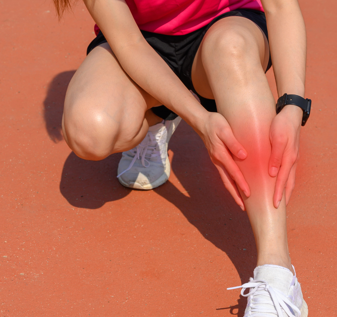
Calf strains are one of the most common injuries we see in running and field sports — but they can happen to anyone.

Whether it’s during sprinting, pushing off quickly, jumping, or even a long walk after doing “too much too soon,” a calf strain can stop you in your tracks.

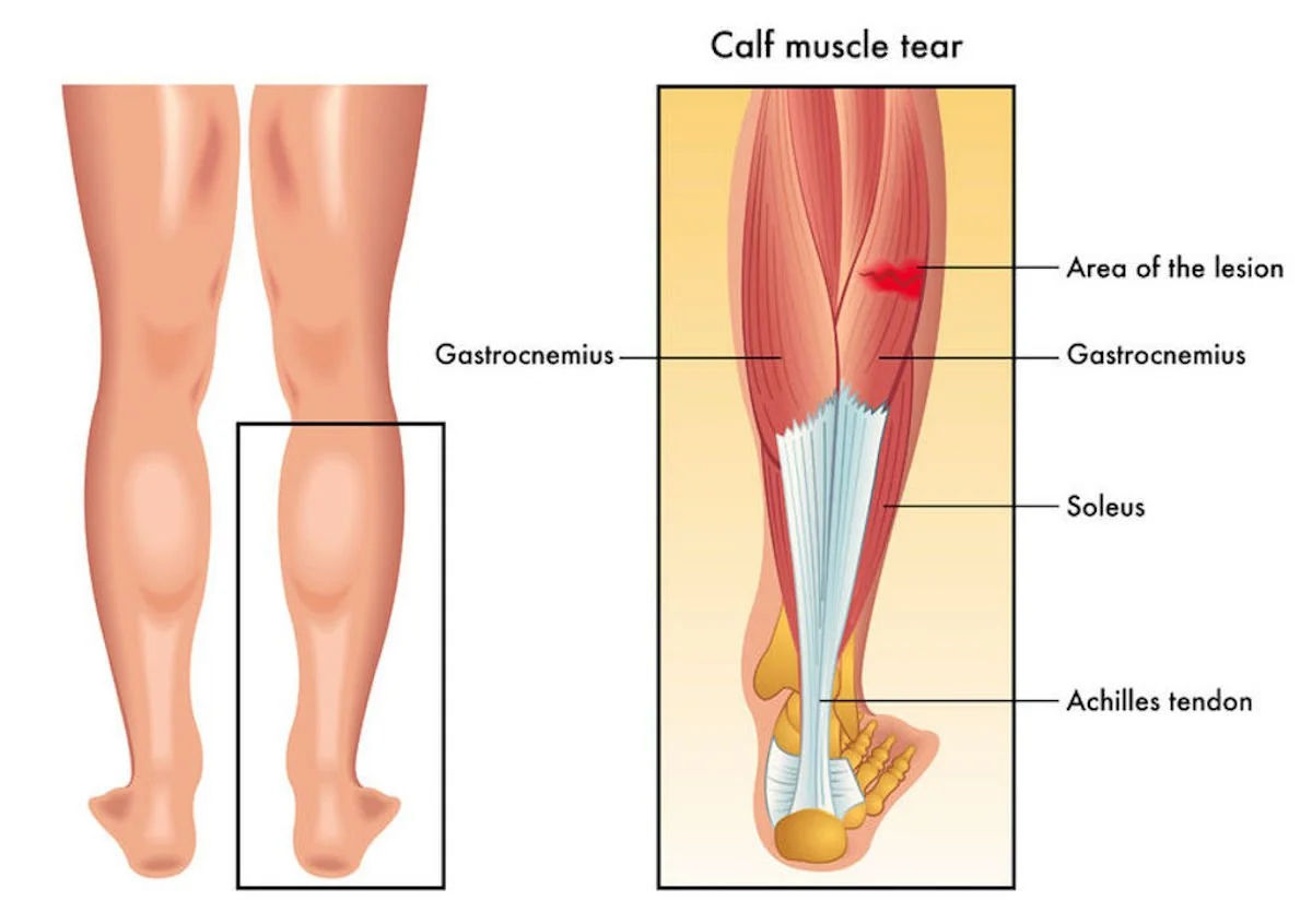
Your calf is made up of two main muscles (the gastrocnemius and soleus) that help you push off, run, jump, and absorb force through the leg. When these muscles are overloaded or stretched too far, small muscle fibres can tear.

What it feels like:

A sudden sharp pain or “pull” in the back of the calf

Tightness or cramping

Pain when walking, pushing off, or going up stairs

Tenderness when touching the muscle

Sometimes swelling or bruising

When it’s worse:

Running or sprinting

Pushing off quickly

Jumping or changing direction

Walking fast uphill or stairs

Why it happens:

Sudden increases in running or sport load

Poor calf strength or endurance

Tight or fatigued muscles

Previous calf injuries

Reduced ankle mobility

How can our Physios help?

Physiotherapists help by reducing pain, restoring strength, and guiding you safely back to sport or activity.

Assessment & diagnosis:
Identify the severity of the strain and what structures are involved

Load management:
Help modify activity so the calf can recover without losing too much fitness

Exercises:
Early movement and strength work

Progressive calf strengthening

Balance and control exercises

Running and return-to-sport progressions

Manual therapy:
Techniques to improve muscle flexibility, ankle mobility, and reduce stiffness

Return-to-sport planning:
Gradually reload the calf safely to reduce the risk of re-injury

Goal:
👉 Reduce pain, rebuild confidence and strength, and get you back to doing what you love as soon as possible.

If you’ve recently injured your calf or keep getting recurring tightness and strains, come see us at the clinic — we’ll help guide your recovery and get you moving again.

Should I see a Physio, Osteo or Chiro?

Physio, Osteo or Chiro? Whats the Difference:

Physiotherapists, Chiropractors, and Osteopaths all treat muscle and joint pain — but they use different approaches.

PHYSIO (What we do well!):

Assessment and diagnosis of injuries and pain

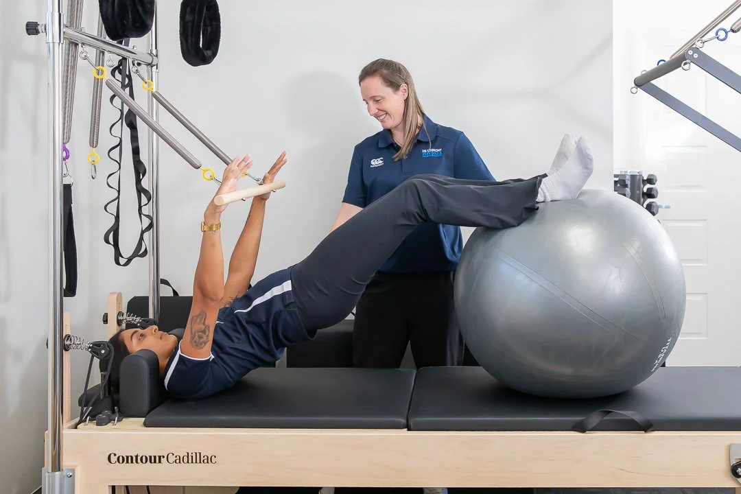
Exercise-based rehab and strengthening

Improving movement, function, and performance

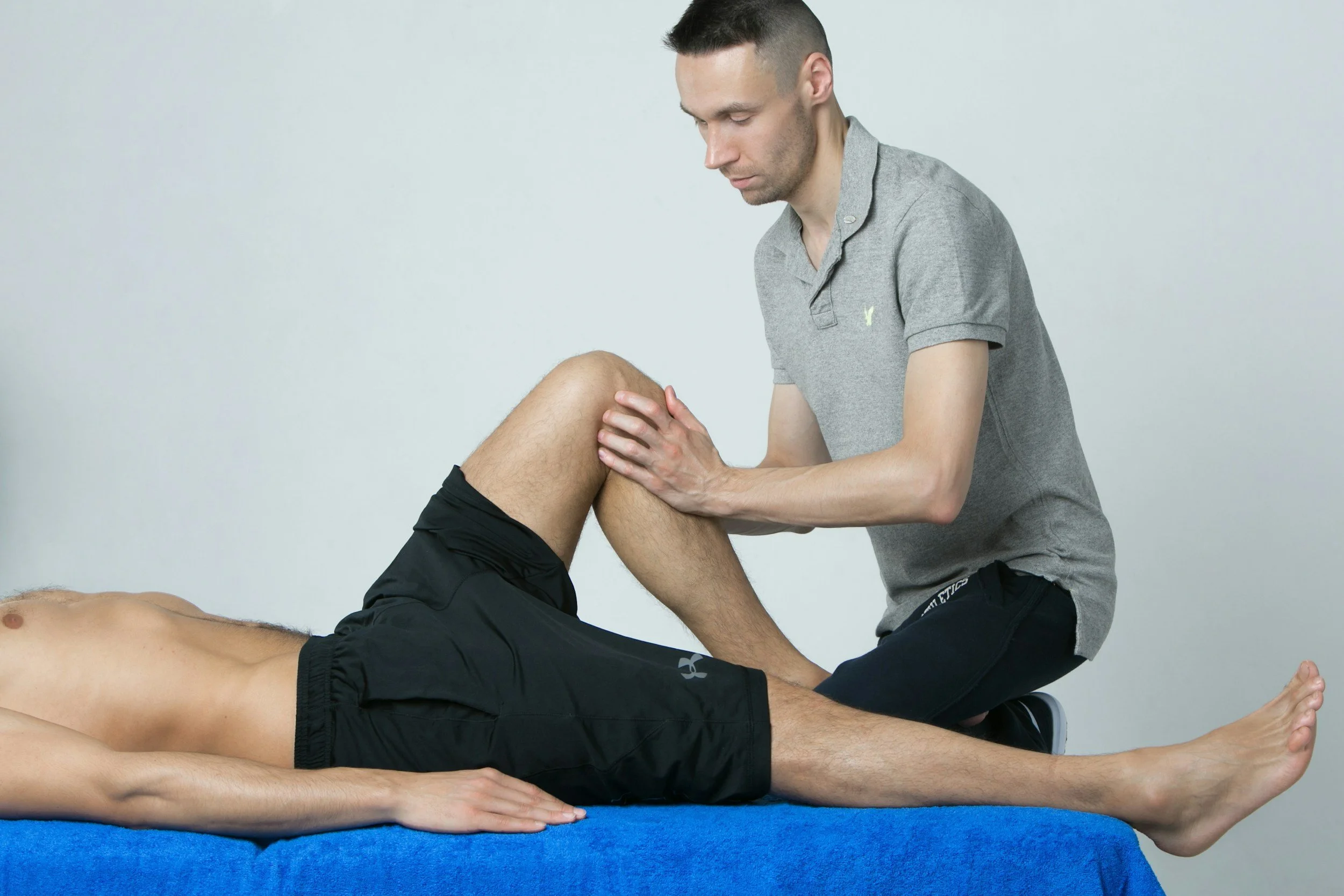
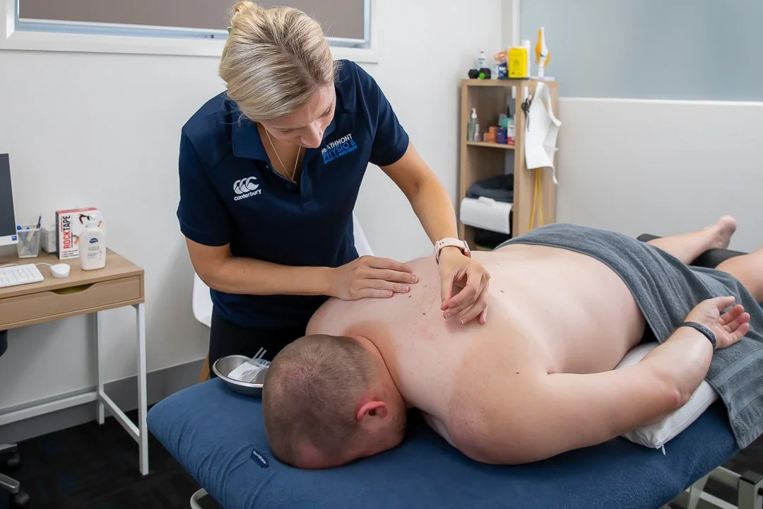
Hands-on treatment including massage, dry needling, cupping and other techniques

Helping you recover and stay active long-term

CHIRO:

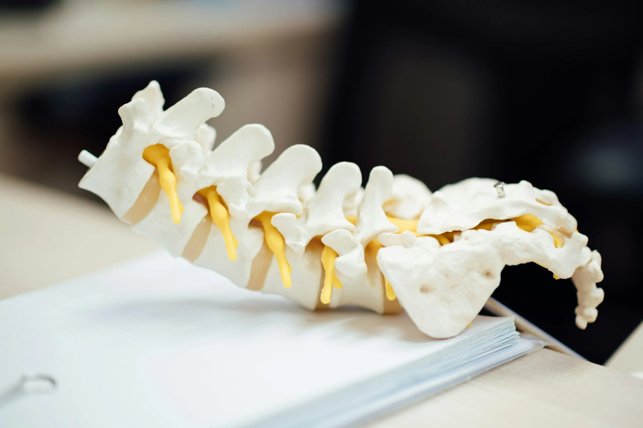
Spinal alignment and joint manipulation

Hands-on treatment of the spine and nervous system

Treatment often centres around “adjustments” or manipulations

OSTEO:

Whole-body movement and alignment

Hands-on manual therapy

Joint mobility, muscles, posture, and movement patterns

SO WHAT MAKES PHYSIOTHERAPY DIFFERENT?

Physiotherapists combine hands-on treatment with exercise rehabilitation and education to help treat the cause of pain — not just the symptoms.

At our clinic, treatment may include:

Exercise programs tailored to your injury and goals

Strength and stability training

Manual therapy and joint mobilisation

Sports rehab and return-to-play planning

Dry needling

Taping and bracing advice

Load management and recovery advice

Post-operative rehabilitation

Education to help prevent flare-ups and future injuries

Physiotherapy is about more than just treating pain — it’s about helping you move better, recover properly, and get back to doing what you enjoy.

Whether you’re dealing with a sports injury, back or neck pain, workplace aches, post-surgery rehab, or ongoing stiffness, our team focuses on finding the cause of the issue and creating a treatment plan tailored to you.

At Heathmont Physio and Sports Medicine, we combine hands-on treatment, exercise rehabilitation, and education to help reduce pain, improve strength and mobility, and prevent injuries from coming back.

👉 If pain or injury is stopping you from performing at your best, come see our team in clinic!

Compartment syndrome

Compartment syndrome is a condition characterised by an increase in pressure within a limited space, this in turn compromises both the circulation and function of the tissues within that space. The space or the compartment consisting of the muscles, blood vessels, arteries and nerves is surrounded by a layer called the fascia. This fascia is a tough connective tissue, hence why it is unable to adapt to increased pressure. Compartment syndrome can be acute onset, or more chronic.


Acute compartment Syndrome:

Acute compartment syndrome is a medical emergency and often presents after a traumatic injury. The most common location for acute compartment syndrome is the anterior or front compartment of the leg that contains the tibialis anterior muscle, extensor muscles of the toes, tibial artery and peroneal nerve. Common conditions that can lead to acute compartment syndrome include:

  • Fractures (Tibial fractures being the most common)

  • Burns

  • Crush injuries

  • Vascular injuries

  • Thrombosis (clots)

  • Bleeding disorders

  • Casts or splints worn incorrectly

  • Tight bandages

  • Intense athletic activity


Symptoms of acute compartment syndrome:

  • Limb pain that is severe

  • Pain not reduced with medication

  • Pain that increases with movement of the limb

  • Burning or tingling

  • Reduced limb strength

  • Temperature reduction

  • Discolouration and swelling (whiteness/ blueness)

  • Reduced mobility of toes and fingers.

Treatment for acute compartment syndrome: A surgery called fasciotomy if often performed, where a cut is made in the skin down to the fascia to release the pressure inside the compartment. This cut remains open to ensure the pressure does not build up again. This surgery aims to decrease the chance of permanent damage to internal structures.

Chronic compartment syndrome:

Chronic or exertional compartment syndrome, unlike acute compartment syndrome is not an emergency. This condition develops over days or weeks and is often an outcome of intense exercise, especially intense activity that is repetitive. The pain usually disappears once the strenuous activity ends.

Symptoms of chronic compartment syndrome:

  • Numbness in the area

  • Tightness in the relevant limb

  • Tingling in the affected area

  • Weakness in the limb

  • Bulge in the muscles

Common causes of chronic compartment syndrome:

  • Enlarged muscles

  • Venous hypertension (increased pressure in veins)

  • Abnormal lack of flexibility in fascia

General treatment of chronic compartment syndrome:

  • Anti-inflammatory Medication

  • Elevation of the affected limb

  • Orthotic shoe inserts

  • Ice post exercise

How can my Physiotherapist help?

Chronic Compartment Syndrome:

  • Diagnosis

  • Adjusting load appropriately

  • Modifying loading biomechanics

  • Addressing muscles imbalances

  • Education on pacing activity

Acute Compartment Syndrome:

  • Diagnosis and acute management including specialist referral

  • Post surgery rehab

  • Preventing stiffness

  • Restoring normal range

  • Muscle training to prevent muscle atrophy

  • Retraining of gait if relevant

  • Retraining for relevant activities (e.g. sports, stairs, occupation)

Compartment syndrome may present similarly to other conditions, hence why it is important to rule out other possibilities.

  • Cellulitis- This condition is a bacterial infection that usually occurs when the skin that acts as a barrier to bacteria is wounded or broken. The swelling, tenderness and tightness of the skin are features that are similar between cellulitis and compartment syndrome, hence why these conditions can be mistaken for each other.

  • Deep Vein Thrombosis- This occurs when a blot clot forms inside a vein, interfering with normal blood flow. Swelling, pain and discoloration are cardinal signs of deep vein thrombosis, and these symptoms as mentioned earlier are also common in compartment syndrome.

  • Gas Gangrene- This is a life-threatening condition caused by a bacterial infection in the muscle leading to rapid tissue destruction and the production of gas. The gas build-up often results in noticeable swelling, hence why gas gangrene can also be mistaken for compartment syndrome.

Take home messages: Compartment syndrome is rare but can have severe complications if diagnosis is late or missed and treatment delayed. If you have had a recent injury and develop severe pain it is important to be seen by your healthcare practitioner as soon as possible for evaluation. If you have been having persistent chronic pain and can’t get to the bottom of its cause, see your physiotherapist for an evaluation and consider chronic compartment syndrome.

References:

McQueen, M. M., & Court-Brown, C. M. (2003). Compartment monitoring in tibial fractures. The pressure threshold for decompression. The Journal of Bone and Joint Surgery. British Volume, 85(5), 693–697. https://doi.org/10.1302/0301-620X.85B5.14352

Agency for Clinical Innovation. (2021). Acute compartment syndrome: Recognition and management of patients with musculoskeletal conditions. NSW Government. https://aci.health.nsw.gov.au/__data/assets/pdf_file/0007/458188/ACI-MSK- Acute-compartment-syndrome.pdf

Kanj, W. W., & Gunderson, M. A. (2022). Acute compartment syndrome. In StatPearls. StatPearls Publishing. https://www.ncbi.nlm.nih.gov/books/NBK448124/

Orthopedic + Sports Medicine Institute of Fort Worth. (n.d.). Compartment syndrome. https://www.osmifw.com/orthopedic-diseases-disorders/compartment-syndrome/

Breathing New Life: The Role of Cardiorespiratory Physiotherapy in Recovery and Wellness

Cardiorespiratory physiotherapy is a specialized area of physical therapy focused on helping individuals with heart and lung conditions improve their physical function, reduce symptoms, and enhance their quality of life. With the increasing prevalence of cardiovascular and respiratory diseases, this form of physiotherapy is becoming more important than ever.

What is Cardiorespiratory Physiotherapy?

Cardiorespiratory physiotherapy involves the assessment and treatment of patients with both acute and chronic heart and lung conditions. This includes diseases such as:

  • Chronic Obstructive Pulmonary Disease (COPD)

  • Asthma

  • Bronchiectasis

  • Pulmonary fibrosis

  • Post-COVID-19 lung complications

  • Heart failure

  • Post-cardiac surgery recovery

Physiotherapists in this field use evidence-based techniques to help restore optimal breathing patterns, improve physical endurance, and educate patients on lifestyle modifications.

Goals of Cardiorespiratory Physiotherapy

  1. Enhance Lung Function
    Through breathing exercises, airway clearance techniques, and positioning, physiotherapists help patients improve ventilation and reduce the work of breathing.

  2. Improve Cardiovascular Fitness
    Tailored exercise programs gradually build endurance and strength, helping the heart pump more efficiently.

  3. Promote Functional Independence
    Helping patients regain their ability to perform daily activities, such as walking, climbing stairs, or simply moving around the house.

  4. Educate and Empower
    Patient education plays a key role in long-term management, especially for chronic conditions. Patients learn about pacing, energy conservation, and how to recognize warning signs.

Common Techniques Used

  • Airway Clearance Techniques (ACTs) such as percussion, postural drainage, and active cycle of breathing.

  • Inspiratory Muscle Training (IMT) to strengthen respiratory muscles.

  • Pulmonary Rehabilitation Programs involving structured exercise and education.

  • Relaxation Techniques to manage breathlessness and anxiety.

  • Standardised Assessment Tools such as the timed up and go, or the 6 minute walk test to compare individuals scores against normative data.

Who Can Benefit?

Anyone recovering from or managing a Cardiorespiratory condition can benefit, including:

  • Patients post-heart surgery or lung surgery

  • Individuals recovering from COVID-19 with persistent respiratory symptoms

  • Seniors experiencing deconditioning or reduced lung capacity

  • People with sedentary lifestyles at risk of cardiovascular disease

Real-World Impact

Pulmonary rehabilitation has been found to significantly reduce frequency and length of hospital admissions for COPD patients and improve quality of life. Similar benefits have been observed in cardiac rehab, which reduces the risk of future cardiac events.

Conclusion

Cardiorespiratory physiotherapy is not just about breathing exercises — it’s about restoring function, improving quality of life, and helping individuals return to the activities they enjoy. If you or a loved one is facing heart or lung challenges, consulting a physiotherapist could be a key step on the road to recovery.

Breathe better. Move better. Live better.


References:

Postel, D., Willekens, M., Werner, S., Hutting, N., & Keesenberg, M. (2022). The effects of reducing the frequency of long-term physiotherapy on patients with severe COPD: a Dutch multicenter study. European Journal of Physiotherapy25(4), 187–192. https://doi.org/10.1080/21679169.2022.2053201

Torres-Sánchez I, Cruz-Ramírez R, Cabrera-Martos I, Díaz-Pelegrina A, Valenza MC. Results of Physiotherapy Treatments in Exacerbations of Chronic Obstructive Pulmonary Disease: A Systematic Review. Physiother Can. 2017;69(2):122-132. doi: 10.3138/ptc.2015-78. PMID: 28539692; PMCID: PMC5435392.

Thomas E, Lotfaliany M, Grace SL, Oldenburg B, Taylor CB, Hare DL, Rangani WT, Dheerasinghe DAF, Cadilhac DA, O'Neil A. Effect of cardiac rehabilitation on 24-month all-cause hospital readmissions: A prospective cohort study. Eur J Cardiovasc Nurs. 2019 Mar;18(3):234-244. doi: 10.1177/1474515118820176. Epub 2018 Dec 14. PMID: 30547678.

How Physiotherapy Helps with Rotator Cuff Pathologies

Shoulder pain can be frustrating, especially when it interferes with your ability to lift, reach, or even sleep comfortably. One of the most common culprits? Rotator cuff pathologies. Whether it's a strain, tendinopathy, or a tear, physiotherapy plays a crucial role in recovery and getting you back to pain-free movement.

What Is the Rotator Cuff?

The rotator cuff is a group of four muscles and their tendons that surround the shoulder joint. These muscles work together to stabilize the shoulder and allow a wide range of motion. Injury to any of these tendons can lead to pain, weakness, and limited function.

Common Rotator Cuff Pathologies

  • Tendinopathy – Degeneration or irritation of the rotator cuff tendons, often due to overuse.

  • Partial or Full-Thickness Tears – Small or complete disruptions in one or more of the tendons.

  • Shoulder Impingement – When the tendons are pinched during shoulder movement.

  • Calcific Tendinitis – Calcium deposits within a tendon causing pain and inflammation.

Signs You Might Have a Rotator Cuff Injury

  • Pain in the shoulder, especially when lifting the arm

  • Weakness or fatigue in the arm

  • Difficulty sleeping on the affected side

  • A clicking or popping sensation with movement

  • Reduced range of motion

  • Bursitis (inflammation of the fluid filled sac within the shoulder capsule)

The Role of Physiotherapy

Physiotherapy is often the first line of treatment and can help most people avoid surgery. A well-structured program can:

  1. Reduce Pain and Inflammation
    Manual therapy, taping, ultrasound, shockwave, dry needling, and modalities like heat or ice can ease pain early in treatment.

  2. Restore Range of Motion
    Gentle mobility exercises help improve shoulder flexibility and prevent stiffness.

  3. Strengthen Supporting Muscles
    A progressive strengthening program targets not only the rotator cuff but also the scapular stabilizers to improve shoulder mechanics.

  4. Improve Posture and Movement Patterns
    Poor posture and faulty mechanics can put extra strain on the rotator cuff. Physiotherapists teach better movement habits that protect your shoulder in daily life.

  5. Support Return to Activity
    Whether you’re an athlete or just want to carry groceries without pain, physiotherapy can guide your safe return to activity with sport- or task-specific training.

When to Seek Help

If shoulder pain persists beyond a few days, is getting worse, or is limiting your ability to do everyday tasks, don’t wait. Early intervention with physiotherapy can lead to quicker recovery and better long-term outcomes.

The Place of Imaging

Around 40% of people aged 30+ will have tears on imaging, which can be asymptomatic. When you see a physiotherapist for shoulder pain, majority of the time imaging won’t be recommended straight away. When imaging has been performed, your physiotherapist will combine your imaging results with the results of the in-person assessment to determine what the main pain driver is. It’s important to not take the imaging report as gospel for shoulders!

Final Thoughts

Rotator cuff injuries don’t have to mean the end of your active lifestyle. With the right physiotherapy approach, you can relieve pain, rebuild strength, and return to the things you love — whether that’s tennis, gardening, or simply getting dressed without pain.


References:

Lawrence RL, Moutzouros V, Bey MJ. Asymptomatic Rotator Cuff Tears. JBJS Rev. 2019 Jun;7(6):e9. doi: 10.2106/JBJS.RVW.18.00149. PMID: 31246863; PMCID: PMC7026731.

Sciarretta FV, Moya D, List K. Current trends in rehabilitation of rotator cuff injuries. SICOT J. 2023;9:14. doi: 10.1051/sicotj/2023011. Epub 2023 May 23. PMID: 37222530; PMCID: PMC10208043.